
Home » Urological Surgery and Treatments » Hydrocele
A hydrocele is defined as the excessive and abnormal accumulation of serous fluid within the tunica vaginalis—a double-layered membrane or sac that surrounds and protects the testicle inside the scrotum. Under normal conditions, there is a minimal amount of fluid between these layers to allow for testicular mobility without friction. However, when there is an imbalance between the production and natural reabsorption of this fluid, it accumulates under pressure, causing a voluminous increase in one or both sides of the scrotal sac.
A hydrocele can appear at any age and is clinically divided into two distinct realities:
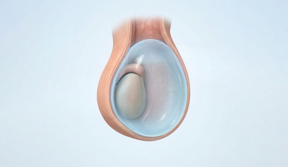
A hydrocele is essentially characterized by being a benign and painless physical alteration. The main symptoms described by patients include:
Diagnosis in a urological office is simple, fast, and painless, essentially aiming to confirm the diagnosis and exclude serious pathologies hidden by the fluid:
Small hydroceles that do not cause physical discomfort, pain, or aesthetic impact do not require any surgical treatment and are kept under surveillance. Medications or ointments have no efficacy in eliminating or reabsorbing this fluid.
When the hydrocele reaches significant dimensions or causes mechanical symptoms, the definitive and recommended treatment is surgical:
Performed in the operating room under general, regional (spinal anesthesia), or local anesthesia with sedation.
Note on Needle Aspiration: Simple drainage or aspiration of the fluid with a needle in the office **is not recommended as a definitive treatment**. It has a recurrence rate (the fluid filling the scrotum again) of practically 100% within a few weeks and carries a high risk of introducing bacteria, causing serious infections or local hemorrhages. It is reserved only for very debilitated patients who cannot undergo any form of anesthesia.
Recovery is usually fast. The patient should use a scrotal support or tight underwear during the first two weeks to minimize natural post-operative swelling. Relative rest is recommended for the first 3 to 5 days, along with local ice application and abstinence from intense physical effort, sports, or sexual activity for about 3 to 4 weeks.
Book your urology appointment with Dr Frederico Furriel, a urologist with consultations in Leiria, Caldas da Rainha and Porto de Mós. Select your preferred location and submit your appointment request.