
Urinary Lithiasis (Kidney Stones)

Urinary Lithiasis (Kidney Stones)

Urinary lithiasis—clinically referred to as renal calculi and commonly known as “kidney stones”—is a condition characterised by the formation of solid, crystalline structures anywhere in the urinary tract (kidneys, ureters, or bladder).

These “stones” develop when urine becomes supersaturated, meaning it contains an excessive concentration of certain salts and minerals and an insufficient amount of fluid to dissolve them. Over time, these minerals clump together, harden, and form crystals that progressively grow.
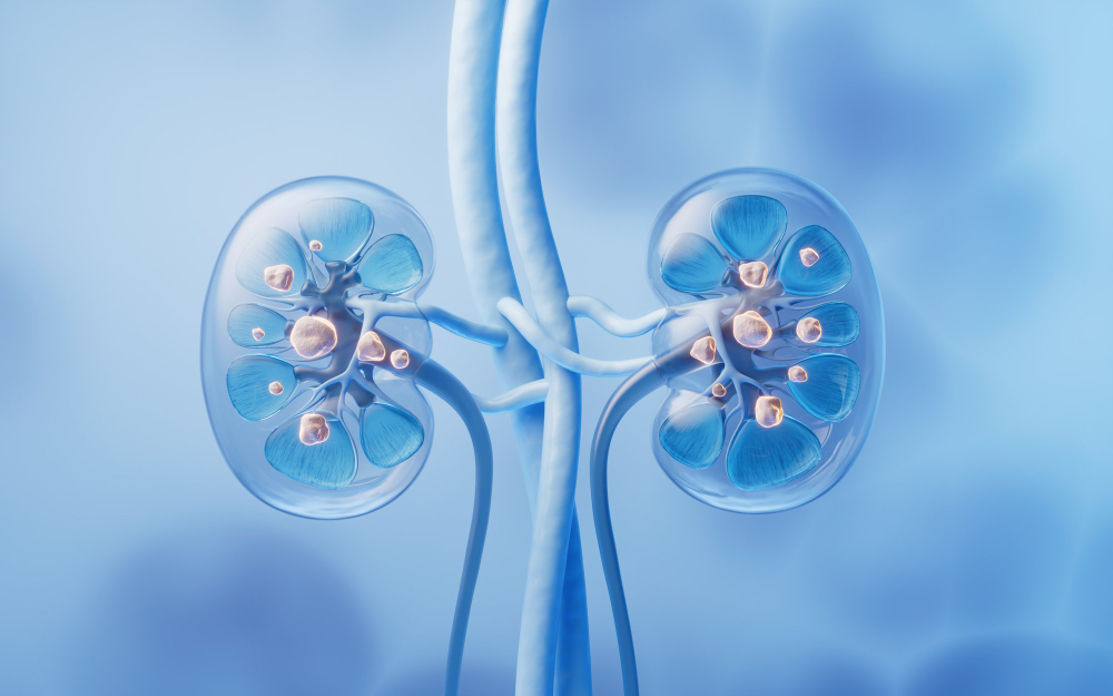
Types of Kidney Stones

The chemical composition of stones varies from patient to patient, depending directly on metabolism and dietary habits:

  • Calcium Stones (Calcium Oxalate and Calcium Phosphate): These are the most common, accounting for around 75% to 80% of cases. They are strongly associated with hereditary and metabolic factors.
  • Uric Acid Stones: More common in men, patients with gout, or those with diets excessively rich in animal protein and organ meats.
  • Struvite Stones (Infection Stones): They result from chronic urinary tract infections caused by specific bacteria. They can grow very quickly, moulding to the cavities of the kidney (staghorn calculi).
  • Cystine Stones: Rare, resulting from an inherited genetic alteration in amino acid transport.
Symptoms of Urinary Lithiasis: From Silent Pain to Renal Colic

A stone may remain inside the kidney for years without causing any symptoms or pain. The real clinical problem arises when the stone moves, attempts to leave the kidney, and becomes lodged in the ureter (the narrow tube that carries urine from the kidney to the bladder), blocking the flow of urine. This causes a sudden, high-pressure dilation of the kidney, triggering the feared renal colic.

The main symptoms include:

  • Severe, Excruciating Flank Pain: A sudden and intense pain on one side of the back, just below the ribs. It is characterised by coming in “waves” (colicky pain), not improving with rest or changes in position, and often radiating to the side of the abdomen, groin, and genitals.
  • Haematuria (Blood in the Urine): The urine may appear pink, reddish, or brownish due to injury caused by the stone passing along the sensitive walls of the urinary tract.
  • Gastrointestinal Symptoms: Severe nausea and vomiting, caused by shared nerve stimulation between the kidney and the digestive system during the pain episode.
  • Urinary Symptoms: A constant, urgent, and painful need to urinate, often passing only a few drops at a time (when the stone is near the bladder).

Warning Signs: When Renal Colic Is an Absolute Emergency

If flank pain or a renal colic episode is accompanied by fever (above 38°C), chills, uncontrollable vomiting that prevents taking oral medication, or complete inability to urinate (anuria), the patient is facing an obstructed and infected kidney. This is one of the most serious urological emergencies, with a risk of progression to urinary sepsis and irreversible loss of kidney function, requiring immediate hospital treatment.

Accurate Diagnosis and Severity Assessment

When lithiasis is suspected or during an episode, the urological diagnosis aims to determine the exact size of the stone, its precise location, and the degree of kidney distress:

  • Computed Tomography (CT / Abdominopelvic CT without contrast): This is the reference test and the most accurate currently available. It can detect virtually 100% of stones (even those not visible on a standard X-ray), measure their density (hardness), and assess in detail the degree of kidney dilation (hydronephrosis).
  • Renal and Bladder Ultrasound: An excellent test for initial screening and monitoring, radiation-free, ideal for assessing kidney size and the presence of dilation.
  • Laboratory Tests (Blood and Urine): Essential to assess overall kidney function (creatinine and urea) and to unequivocally rule out an active urinary tract infection through leukocyturia or a positive urine culture.
Treatments and Surgeries for Urinary Lithiasis

The therapeutic approach to urinary lithiasis depends entirely on two fundamental factors: the size of the stone and its exact anatomical location. A stone lodged in the ureter requires a completely different strategy from a stone that is stable within the kidney.

A. Treatment of Ureteral Stones (Stones in the Urinary Channel)

When a stone leaves the kidney and becomes lodged in the ureter, it blocks the normal flow of urine and triggers a renal colic episode. Here, the main goal is to relieve the obstruction quickly and safely:

  • Medical Expulsive Therapy (MET): This clinical approach applies exclusively to stones located in the ureter, that are small (generally less than 4 to 5 mm) and are not causing complications (such as fever or kidney deterioration). It consists of combining analgesics and potent anti-inflammatory drugs to control pain, together with medications that relax the muscles of the urinary tract (alpha-blockers, such as tamsulosin), to facilitate and speed up the spontaneous passage of the stone in the urine.
  • Semi-Rigid Ureterorenoscopy with Laser: This is the surgical treatment of choice for the vast majority of ureteral stones (especially in the mid and distal ureter, closer to the bladder), or when medical treatment fails. Without any skin incision, the urologist inserts a thin, firm instrument with a high-definition camera (semi-rigid ureteroscope) through the urethra, advancing up the ureter to the exact site where the stone is lodged. The stone is fragmented with a laser (Holmium or Thulium) fibre, and the pieces are immediately removed with small basket-shaped forceps (baskets).
  • Extracorporeal Shock Wave Lithotripsy (ESWL): It may be considered a non-invasive alternative for small stones located in the upper ureter (closer to the kidney), using focused acoustic waves from outside the body to try to break the stone without the need for anaesthesia.

B. Treatment of Renal Stones (Stones Inside the Kidney)

For stones that are lodged and stationary within the cavities of the kidney itself, the strategy is based mainly on stone size, the presence of symptoms, and the risk of future complications:

  • Active Surveillance (Monitoring): Not all kidney stones need to be treated or operated on immediately. If the patient has small stones (generally under 5–6 mm), located in renal calyces that do not cause obstruction, and the situation is completely asymptomatic (no pain or infections), the recommended approach is clinical surveillance. This consists of periodic imaging tests (such as ultrasound or X-ray) and regular appointments to monitor whether the stone remains the same size or shows signs of progression, thereby avoiding surgical interventions.
  • Flexible Ureterorenoscopy / RIRS (Retrograde Intrarenal Surgery): This is the modern gold-standard treatment for moderate-sized kidney stones (generally up to 2 cm) that require intervention (because they cause pain, recurrent infections, or are growing). It uses a highly manoeuvrable, flexible digital endoscope that navigates the urinary tract to reach the inside of the kidney, accessing the deepest renal calyces without any skin incision. The urologist uses state-of-the-art laser technology to dust the stone or break it into millimetre-sized fragments for subsequent removal.
  • Percutaneous Nephrolithotomy (PNL / PCNL): This is the standard reference surgery indicated for large, complex kidney stones or stones larger than 2 cm (including staghorn calculi, which occupy the entire internal cavities of the kidney). Under general anaesthesia, a small incision of only 1 cm is made in the lumbar (back) region, creating a direct tract to the kidney that allows the insertion of a camera (nephroscope). Using powerful energy systems, the large stone is fragmented and aspirated out very effectively, with an average hospital stay of only 2 to 4 days.
  • Extracorporeal Shock Wave Lithotripsy (ESWL): It remains indicated for small kidney stones (<1–1.5 cm) that require treatment, located in the upper or mid calyces of the kidney and with moderate density (less hard stones), as a fully outpatient approach.
Special FAQ Section: Methods for Relieving Obstruction and Draining the Kidney

These are essential procedures to prevent or resolve acute urinary blockages, preserve kidney function, and ensure patient safety in the post-operative period.

The Double-J (JJ) Ureteral Catheter / Stent

The Double-J catheter is an extremely thin, flexible, biocompatible plastic tube. Its ends are curled (in a spiral or “pigtail” shape) to secure one end inside the kidney and the other inside the bladder, preventing the catheter from moving. It remains completely hidden inside the body.

  • What is it for? It works as an internal “bridge” or alternative channel. It ensures the ureter stays open and that urine can flow freely from the kidney to the bladder, even if the channel is very inflamed due to trauma from a stone passing or after endoscopic laser surgery.
  • How is it inserted and removed? It is inserted endoscopically through the urethra in the operating theatre. Removal is a simple and very quick procedure (seconds), performed in the urology clinic under local anaesthetic gel via flexible cystoscopy.
  • What is normal to feel (Expected Symptoms): While the patient has a Double-J catheter in place, it is perfectly normal and common to experience increased urinary frequency, a sense of urgency to urinate, mild back (kidney) discomfort at the exact moment of urination, and small streaks of blood in the urine (haematuria), especially after physical exertion or walking. These symptoms are normal, benign, and resolve completely and immediately once the catheter is removed from the body.

Percutaneous Nephrostomy

When the kidney is severely obstructed, very dilated, and has an active infection (accumulation of pus or infected urine under pressure), and it is not technically possible or safe to bypass the blockage endoscopically through the bladder, the immediate solution to save the patient’s life and the kidney is a percutaneous nephrostomy.

  • The Procedure: This is a rapid, emergency intervention performed under local anaesthesia and guided by real-time ultrasound or X-ray. The urologist inserts a thin drainage catheter through the skin of the back directly into the dilated kidney.
  • Vital Function: It allows infected urine under pressure to be drained immediately into an external collection bag attached to the body. This provides immediate pain relief, drains the infection, and protects the kidney from severe and permanent damage. Definitive treatment of the stone that caused the obstruction is safely postponed to a second stage, once the patient is clinically stable and free of infection.


Special Note: Bladder Stones (Stones in the Bladder)

Although they are part of urinary lithiasis, bladder stones have a completely different origin and clinical approach from stones that form in the kidneys.

  • The Underlying Cause: Only rarely does a bladder stone result from a stone that has descended from the kidney. In the vast majority of cases, these stones form directly inside the bladder itself due to bladder outlet obstruction—that is, a blockage at the urine outflow. The most common cause is an enlarged prostate (Benign Prostatic Hyperplasia), which prevents complete emptying of the organ. Urine that is chronically retained and accumulated in the bladder (post-void residual urine) eventually stagnates, becomes concentrated, and crystallises, giving rise to bladder stones.
  • Surgical Treatment (Laser Cystolithotripsy): Bladder stones are not passed spontaneously and almost always require surgical treatment, which today is performed in a fully minimally invasive manner. The standard procedure is Endoscopic Laser Cystolithotripsy. Without any skin incision, the urologist inserts a camera (cystoscope) through the urethra into the bladder. Using a high-power laser fibre, the stone is fragmented and reduced to dust or small grains, which are then aspirated and washed out.

Combined Approach: As the bladder stone is only a symptom of a larger problem, it is very common to use the same surgical time and the same anaesthesia to treat the cause of the obstruction (for example, by simultaneously performing prostate surgery, such as TURP, laser, Aquablation, or simple prostatectomy), preventing new stones from forming again in the future.

Have you been diagnosed with, or do you have a history of, kidney stones?
Urinary lithiasis requires a specialised and timely approach to prevent painful episodes and protect your kidney function. Have all your questions about treatments and post-operative care answered.

Appointment booking

Book your urology appointment with Dr Frederico Furriel, a urologist with consultations in Leiria, Caldas da Rainha and Porto de Mós. Select your preferred location and submit your appointment request.